
Retinal Detachment: A Sight-Threatening Emergency You Shouldn’t Ignore
- visionopolisclinic
- Oct 25, 2025
- 3 min read

Updated: Dec 25, 2025
# Understanding Retinal Detachment: A Vision-Threatening Emergency
## What is Retinal Detachment?
Retinal detachment occurs when the retina pulls away from the layer of blood vessels that supplies it with oxygen and nutrients. Without prompt treatment, this separation can lead to permanent vision loss. This is not a condition that improves on its own; it requires urgent medical intervention from a retinal specialist.

## Types of Retinal Detachment

There are three main types of retinal detachment:
Rhegmatogenous Retinal Detachment (most common): This type is caused by a tear or hole in the retina, allowing fluid to pass underneath and separate the retina from the underlying tissue.
Tractional Retinal Detachment: This occurs when scar tissue on the retina’s surface contracts and pulls the retina away. It is often seen in advanced diabetic retinopathy.
Exudative Retinal Detachment: This type is caused by inflammation, injury, or tumors that lead to fluid buildup under the retina without any tear.
## Symptoms: Warning Signs You Should Never Ignore

Retinal detachment is often painless, but it comes with sudden and alarming changes in vision. Be aware of the following symptoms:
⚠️ Sudden appearance of floaters (tiny dark spots or threads)
⚠️ Flashes of light in one or both eyes
⚠️ A shadow or curtain falling over your field of vision
⚠️ Blurred or distorted vision
⚠️ Sudden decrease in central or peripheral vision
These symptoms may come and go quickly. Don’t wait. Seek emergency eye care immediately.
## Causes and Risk Factors
Several factors can increase the risk of retinal detachment:
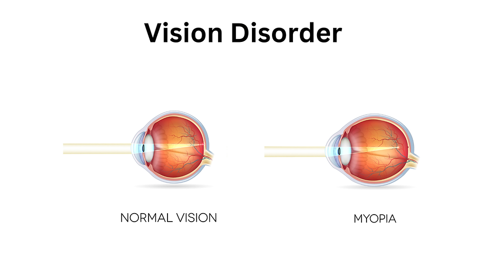
High myopia (nearsightedness)
Aging (most common in people over 50)
Eye trauma or injury
Previous eye surgery, such as cataract removal
Family history of retinal detachment
Diabetic retinopathy or other retinal diseases
Understanding these risk factors can help in early detection and prevention.
## Diagnosis: How Eye Specialists Detect Retinal Detachment
Eye specialists use specialized tools for early and accurate diagnosis. Some of these tools include:
Dilated Fundus Exam: This is a comprehensive retinal evaluation using high-powered lenses.
Optical Coherence Tomography (OCT): This produces detailed cross-sectional images of the retina.
Ultrasound Imaging (B-scan): This is useful when the retina isn’t clearly visible due to bleeding or clouding.
## Treatment Options for Retinal Detachment
Retinal detachment requires urgent surgical repair. The treatment approach depends on the type, location, and extent of the detachment. Here are some common options:
🔹 Laser Photocoagulation or Cryopexy
This is used in early stages (before detachment) to seal retinal tears.
🔹 Pneumatic Retinopexy
In this procedure, a gas bubble is injected into the eye to push the retina back in place. This is often combined with laser or freezing treatment.
🔹 Scleral Buckling
A flexible band is placed around the eye to counteract the pull of the retina and help it reattach.
🔹 Vitrectomy
This involves the removal of the vitreous gel to allow access to the retina and repair tears. It is often used in complex cases.
## Prognosis: Can Vision Be Restored?
Timely treatment offers the best chance of saving your sight. If the macula (the central part of the retina) is still attached, vision can often be preserved. However, delayed treatment may result in permanent vision loss, especially if the macula detaches.
Follow-up care is essential. This includes avoiding strenuous activity, maintaining proper head positioning (if advised), and adhering to all post-operative guidelines.
## Prevention: How to Protect Your Retina
While not all cases are preventable, you can reduce your risk by:
🧿 Managing high myopia and diabetes
🧿 Wearing protective eyewear during sports or hazardous work
🧿 Getting regular dilated eye exams, especially if you're at high risk
🧿 Reporting any vision changes promptly
## Final Thoughts: Your Vision Is Too Valuable to Risk
Retinal detachment is a vision-threatening emergency. However, with prompt diagnosis and treatment, it is often possible to restore or preserve sight. At our hospital, we’re equipped with advanced diagnostic tools and surgical expertise to handle all retinal emergencies with precision and care.
If you're experiencing sudden vision changes, don’t delay. Schedule an emergency consultation with your eye specialist immediately. Your vision is too valuable to risk.


Excellent article that balances medical accuracy with reader-friendly language. The section on risk factors and treatment options helps remove confusion and encourages timely action. Awareness is key when it comes to retinal health, and resources like this are extremely valuable. It also complements the education provided through professional Retinal Care in Draper services and eye health programs.
The academic framework at reading comprehension for class 3 highlights clarity and applied understanding. Learning content is informed by industry practice. Academic precision supports practical execution. Flexible scheduling accommodates varied routines. This keeps learning manageable.
The academic framework at Class 5 CBSE English highlights clarity and applied understanding. Learning content is informed by industry practice. Academic precision supports practical execution. Flexible scheduling accommodates varied routines. This keeps learning manageable.
The academic framework at UNICCM School highlights clarity and applied understanding. Learning content is informed by industry practice. Academic precision supports practical execution. Flexible scheduling accommodates varied routines. This keeps learning manageable.
यह जानकर बहुत चिंता होती है कि रेटिनल डिटैचमेंट एक गंभीर आपात स्थिति है जिसे नजरअंदाज नहीं किया जाना चाहिए; क्या आप बता सकते हैं कि सर्जरी के बाद दृष्टि (vision) को पूरी तरह से ठीक होने में आमतौर पर कितना समय लगता है? सादर प्रणाम <a href="https://jakarta.telkomuniversity.ac.id/belajar-gagal-bangkit-siklus-normal-kehidupan-mahasiswa/">Telkom University Jakarta</a>